
Background
In planar lipid bilayers and molecular models, electropores emerge in nanoseconds once the critical TMP is reached and collapse within microseconds after PEF. In stark contrast, live cells remain permeabilized for minutes or even hours. This prolonged permeabilized state enables all electroporation-based therapies and also underlies their adverse side effects. No existing electroporation theory adequately explains this persistent membrane leakiness. Limited understanding of the electrical stress response mechanisms impedes the advancement of existing PEF-based therapies. Primary membrane effects of PEF determine diverse downstream physiological effects and cell survival. A detailed understanding of these early events is key to developing effective treatments and minimizing adverse effects.
Our team has recently advanced the field by dynamic imaging of single electropores, measuring their conductance, and capturing cell membrane charging and relaxation kinetics on the nanosecond time scale. This project will explore membrane response to PEF at the single-pore level and with nanosecond resolution, to establish the primary mechanisms of complex electropermeabilization phenomena and their dependence upon PEF parameters and protocols.
Aim and Objects
Aim 1: Quantify the formation of membrane lesions (diffuse permeabilization, transient and persistent focal pores) with respect to PEF duration (nano- to milliseconds) and the electric field strength (0.1-10 kV/cm). Compare effects of single PEFs and PEF trains at 10–90% duty cycles. Correlate lesion types with membrane charging and relaxation kinetics in cells of different shape and excitability. Characterize membrane charging by MHz compression of nsPEF bursts and bipolar cancellation mechanisms.
Aim 2: Characterize the lifecycle, permeability, and current-voltage function of single electropores and their dependence on cell physiology, environment, and PEF protocols. Investigate electropore association with lipid rafts, voltage-gated channels, and structural proteins. Analyze whether membrane proteins form electropores and/or contribute to electropore structure and longevity. Test the electrodeformation mechanism of electropermeabilization.
Aim 3. Examine the protective role of the adaptive electropore conductance and analyze the underlying mechanisms. Design novel nsPEF protocols to control electropermeabilization. Quantify cellular protection conferred by the activation of voltage-gated ion channels, lanthanide ions, and poloxamers.
Expected outcomes
Understanding the kinetics of membrane charging and relaxation, diverse injury mechanisms, their interaction and dependence on PEF parameters and cell physiology will create a logical framework for fine tuning PEF effects for either efficient stimulation without damage, or towards efficient ablation with minimal neuromuscular stimulation. New mechanistic knowledge will create the theoretical basis for novel medical treatments.
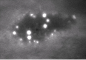
Figure legend: Ca2+ “fountains” into cell cytosol through electropores made visible by TIRF microscopy
Project details
| Principal investigator | Andrei Pakhomov |
|---|---|
| Role | PI |
| Home institution | Frank Reidy Research Center for Bioelectrics Old Dominion University |
| Funder | NIH/NIGMS |
| Start date | June 2026 |
| End date | March 2030 |
| Grant budget | $1,280,000 |
| Participant institutions | Old Dominion University, USA |